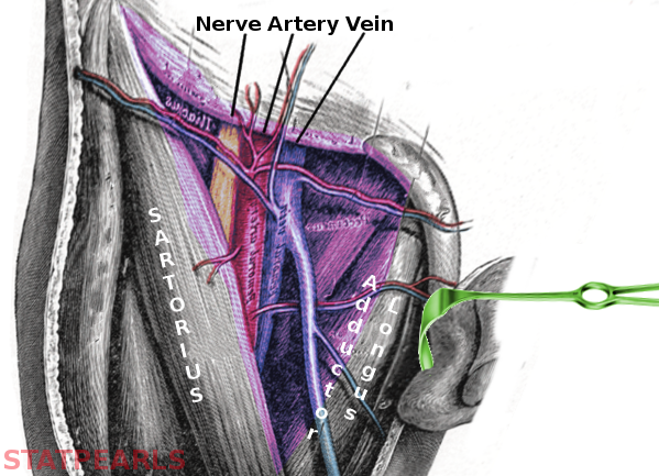
Anatomy Of The Femoral Nerve Artery And Vein Medical Click Images to Large View Anatomy Of The Femoral Nerve Artery And Vein Medical Illustration Of The Femoral Nerve Block Region Showing The Click Images to Large View Illustration Of The Femoral Nerve Block Region Showing The Femoral Nerve Anatomy And Clinical Notes Kenhub Click Images to Large View Femoral Nerve AnatomyHere are the femoral artery and vein at the point where we saw them last, disappearing beneath the sartorius muscle To follow their course, we'll remove sartorius, and also gracilis Here's vastus medialis, here's adductor longus, with adductor magnus behind it The femoral vessels pass beneath the roof of the adductor canal, and through the adductor hiatus To see where they The saphenous nerve, artery, and vein are integral structures of a neurovascular bundle that courses through the thigh and leg of the lower limb Firstly, the saphenous nerve is a strictly sensory nerve with no motor function It is responsible for innervation to the anteromedial aspect of the leg The saphenous artery, a distant branch of the femoral artery arising from the
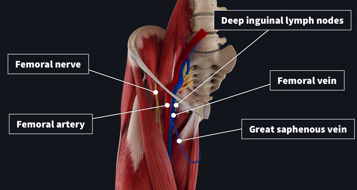
Remember The Contents Of The Femoral Triangle With This Crafty Mnemonic Complete Anatomy
Femoral artery vein nerve anatomy
Femoral artery vein nerve anatomy-Its contents are shown below (from lateral to medial) Femoral branch of the genitofemoral nerve occupies the lateral compartment of the femoral sheath along with femoral Femoral artery and its branches It emerges from the base of the femoral triangle at the midinguinal point (midpointFemoral nerve â€" innervates the anterior compartment of the thigh, and provides sensory branches for the leg and foot Femoral artery â€" responsible for the majority of the arterial supply to the lower limb Femoral vein â€" the great saphenous vein drains into the femoral vein


Femoral Triangle
At the inguinal ligament it becomes the external iliac vein;Find it here, along with the essential anatomy you need to know about this artery Looking for a mnemonic to remember the branches of the femoral artery?Femoral_anatomy_vein_artery_nerve 1/3 Femoral Anatomy Vein Artery Nerve Download Femoral Anatomy Vein Artery Nerve Internal Intraabdominal HerniasRoberto L Estrada 1994 Atlas of Surgical Techniques in TraumaDemetrios Demetriades Hundreds of highquality intraoperative photos of fresh human cadavers create a uniquely realistic stepbystep
Summary origin continuation of the superficial femoral artery as it exits the adductor canal main branch anterior tibial artery termination continues as the tibioperoneal trunk in the lower aspect of the popliteal fossa supply knee, leg and foot Gross anatomy Origin As a continuation of the femoral (superficial femoral) artery as it passes into the popliteal fossa through the adductor This artery crosses the femoral nerve and femoral vein in such a way as to form a delta shape near the groin region This portion is known as the femoral triangle or Scarpa's triangleFEMORAL TRIANGLE superior inguinal ligament;
The femoral nerve (also anterior crural nerve, latin nervus femoralis) is the largest nerve of the lumbar plexus, which arises from the anterior rami of the first, second, third and fourth lumbar nerves (L1 L4) The femoral nerve is a mixed nerve containing motor and sensory fibersRelevant Anatomy for Femoral Artery Cannulation, USGuided The femoral artery and vein are accessible within the femoral triangle, which is defined by the inguinal ligament superiorly, the adductor longus muscle medially, and the sartorius muscle laterally The inguinal ligament is defined as a line drawn between the symphysis pubis and the anterior superior iliac spine The femoral arteryThe saphenous nerve, artery, and vein are integral structures of a neurovascular bundle that courses through the thigh and leg of the lower limb Firstly, the saphenous nerve is a strictly sensory nerve with no motor function1 It is responsible for innervation to the anteromedial aspect of the leg The saphenous artery, a distant branch of the femoral artery arising from the


Femoral Triangle


Illustration Of The Femoral Nerve Block Region Showing The Femoral Download Scientific Diagram
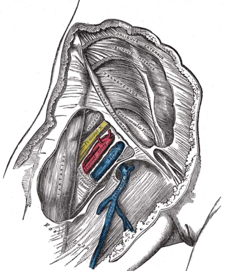
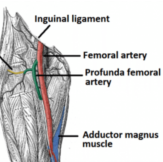
Mnemonics to recall the order of the femoral vessels and nerve as they emerge from beneath the inguinal ligament into the femoral triangle are NAVY; The femoral artery, vein, and nerve all exist in the anterior region of the thigh known as the femoral triangle, just inferior to the inguinal ligament Within the femoral triangle, the anatomical relationship from medial to lateral is femoral vein, common femoral artery, and femoral nerve The artery and vein are both contained within the femoral sheath while the nerveN = femoral nerve A = femoral artery V = femoral vein EL = empty space (femoral canal) and lymphatics The deep femoral artery arises in the femoral triangle It passes deep to the adductor longus and gives rise to perforating arteries that supply the posterior thigh The medial and lateral femoral circumflex arteries are typically branches of the deep femoral artery The medial
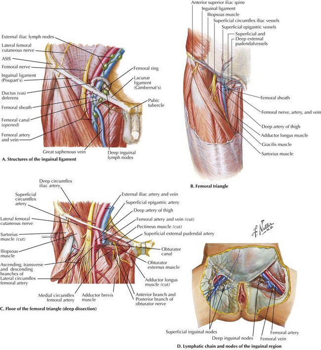


Exposure Of The Common Femoral Artery And Vein Basicmedical Key


Section 2 Anatomy And Physiology
The femoral branch of the genitofemoral nerve is also lateral to the upper part of the femoral artery, within the femoral sheath, but lower down it passes to the front of the artery • 6 The profunda femoris artery a branch of the femoral artery itself, and its companion vein, lie behind the upper part of the femoral artery, where it lies on the pectineus – Lower down,Anatomy_of_femoral_artery_and_vein 3/18 Anatomy Of Femoral Artery And Vein Circulation through the deep femoral artery and its branches is critical to patients with aortoiliac and infrainguinal arteriosclerosis It is, accordingly, essential that all physicians who are seriously interested in treating patients with lower extremity ischemia have a good working knowledge of this crucial artery After arising from the lumbar plexus, the femoral nerve travels inferiorly through the psoas major muscle of the posterior abdominal wall It supplies branches to the iliacus and pectineus muscles prior to entering the thigh The femoral nerve then passes underneath the inguinal ligament to enter the femoral triangle Within this triangle, the nerve is located lateral to the femoral vessels (unlike the nerve, the femoral artery and vein are enclosed within the femoral


Neurovasculature Of The Lower Limbs Knowledge Amboss


Femoral Nerve Origin Course Branches Applied Anatomy
Y "Yfronts" (ie the midline) The terminal cutaneous branch of the femoral nerve is the saphenous nerve It travels through the adductor canal (accompanied by the femoral artery and vein) and exits prior to the adductor hiatus The saphenous nerve innervates the medial aspect of the leg and the footPopliteal Vein, lateral Superior Genicular Artery, deep Artery Of The Thigh, popliteal Artery, femoral Artery, Muscular system, artery, blood Vessel, nerve, human Anatomy lateral Compartment Of Leg, sural Nerve, common Peroneal Nerve, deep Vein Of The Thigh, lateral Cutaneous Nerve Of Thigh, peroneus Longus, crus, vein, blood Vessel, nerve


Remember The Contents Of The Femoral Triangle With This Crafty Mnemonic Complete Anatomy


Precautions For Manual Therapy Of The Lumbar Spine And Pelvis
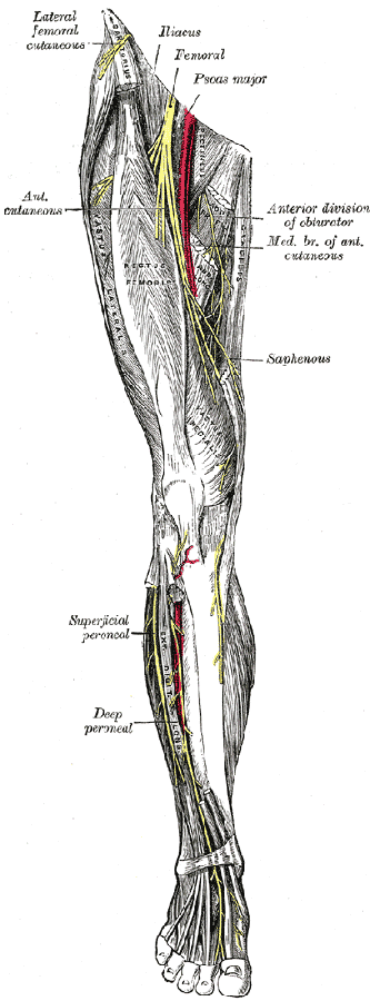
The femoral nerve combines nerve fibers that emerge from between the second, third, and fourth lumbar (lower back) vertebrae As it extends downward, it branches off to the skin, muscles, and connective tissues of the hip and thigh, including the iliacus muscle (a thigh flexor) and the inguinal ligament (in the groin)The femoral nerve is located lateral to the femoral artery, outside the femoral sheath, in the groove between the iliacus and the psoas major About 25 cm below the inguinal ligament it divides into anterior and posterior sections which enclose lateral circumflex femoral artery between them The anterior section produces 2 cutaneous branches intermediate and medial cutaneous nervesSaphenous nerve The saphenous nerve (also long saphenous nerve, internal saphenous nerve, latin nervus saphenus) is a large cutaneous branch of the femoral nerve The saphenous nerve contains only sensory fibers The saphenous nerve runs posterior to the sartorius, enters the adductor canal and pierces the anterior wall of the channel


Anatomy Lectures Femoral Artery Femoral Vein Femoral Nerve Youtube
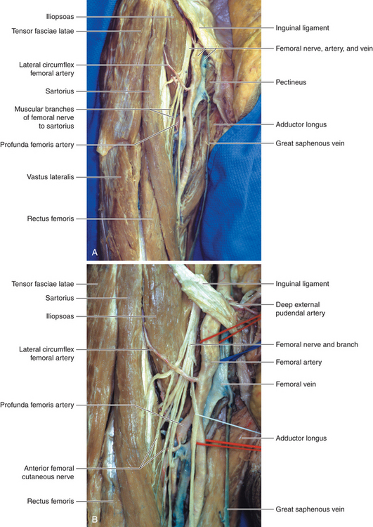


Femoral Nerve Neupsy Key
The proximal femoral artery and vein are wrapped in a fibrous covering called the femoral sheath This sheath is made up of several components The lateral part of the sheath adjacent to the femoral nerve is the continuation of the iliac fascia covering the iliopsoas muscle The posterior portion of the sheath is the fascia covering the pectineus muscle Anteriorly andI am so proud to present you today this article, that maThe femoral artery is a large vessel that provides oxygenated blood to lower extremity structures and in part to the anterior abdominal wall The common femoral artery arises as a continuation of the external iliac artery after it passes under the inguinal ligament The femoral artery, vein, and nerve all exist in the anterior region of the thigh known as the femoral triangle, just inferior to


Www Medicinebau Com Uploads 7 9 0 4 19 Muscles Of Thigh Pdf


Ultrasound Guided Saphenous Adductor Canal Nerve Block Nysora
Adductor Magnus Anatomy Medbullets Step 1 Hello, What's up guys?The femoral artery vein and nerve all exist in the anterior region of the thigh known as the femoral triangle just inferior to the inguinal ligament The primary function of this artery is to supply blood to the lower section of the body Femoral Artery Orthopaedicsone Articles Orthopaedicsone The femoral artery is a large artery in the thigh and the main arterial supply to the thigh and legThe femoral branch of the genitofemoral nerve is also lateral to the upper part of the femoral artery, within the femoral sheath, but lower down it passes to the front of the artery 6 The profunda femoris artery a branch of the femoral artery itself, and its companion vein, lie behind the upper part of the femoral artery, where it lies on the pectineus


Femoral Vein Radiology Reference Article Radiopaedia Org


Us Guided Regional Anesthesia Archives Page 45 Of 53 Usabcd
Occipital Artery, greater Occipital Nerve, supraorbital Artery, superficial Temporal Artery, supraorbital Nerve, facial Nerve, head And Neck Anatomy, artery, scalp, nerve popliteal Vein, lateral Superior Genicular Artery, deep Artery Of The Thigh, popliteal Artery, femoral Artery, Muscular system, artery, blood Vessel, nerve, human AnatomyProfessor, Department Chair, Surgeon, Neuroscientist and Medical Informatician in the Western HemisphereIt sStudy Femoral sheath, artery, vein and nerve (dave's notes) flashcards from Janet Rhodes's class online, or in Brainscape's iPhone or Android app Learn faster with spaced repetition


Section 5 Ultrasound Theory And Point Of Care Application


Femoral Artery Femoral Nerve Arteries Mnemonics
In the majority of cases (modal anatomy), the femoral vein is the main vessel, but it could be the axial vein or the deepfemoral vein, or it could be separated into two main trunks All these can be considered as truncular venous malformations, occurring at a late stage of the embryonic development 6 Material and methods This study is an update of an initial studyMedial border adductor longus;Accompanies the femoral artery in the femoral triangle;


Http Ksumsc Com Download Center Archive 1st 439 2 muscloskeletal block Team work Anatomy 18 vascular anatomy of the lower limb Pdf
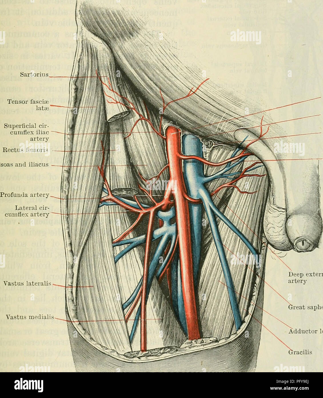


Cunningham S Text Book Of Anatomy Anatomy The Deep Veins Of The Lowek Extremity 987 The Medial Side Of The Femoral Artery About 37 Mm One And A Half Inches Below The Inguinal
The femoral artery is a large artery in the thigh and the main arterial supply to the thigh and leg The femoral artery gives off the deep femoral artery or profunda femoris artery and descends along the anteromedial part of the thigh in the femoral triangle It enters and passes through the adductor canal, and becomes the popliteal artery as it passes through the adductor hiatus in the adductor Femoral triangle anatomy The neurovascular bundle consists of the femoral vein, artery, and nerve, which lie within the triangle in that order from medial to lateral The femoral sheath enclosesFind it here, along with the essential anatomy you need to know about this artery Today Explore When the autocomplete results are available, use the up and down arrows to review and Enter to select


S F Physical Exam Flashcards Quizlet


The Femoral Triangle And Superficial Veins Of The Leg Anaesthesia Intensive Care Medicine
ANATOMY • COMMON femoral vein in groin • Formed by union of deep and superficial femoral veins • Becomes External Iliac vein at inguinal ligament ANATOMY • Great saphenous vein – enters approx 3cm below inguinal ligament – good landmark on US • Medial and lateral circumflex veins ANATOMY • Vein lies in femoral triangle with • femoral nerve • femoral artery • lymphatics Femoral Vein Anatomy continuation of the popliteal vein;N = femoral nerve A = femoral artery V = femoral vein EL = empty space (femoral canal) and lymphatics The femoral nerve provides motor innervation to the muscles of the anterior compartment of the thigh, with a few exceptions the psoas portion of iliopsoas is innervated by muscular branches of the lumbar plexustensor fasciae latae is innervated by the superior gluteal nerve


Dissector Answers Anterior Medial Thigh


Total Hip Replacement Doctor Stock
The anterior femoral cutaneous vein carries oxygendepleted blood from the capillaries of the superficial anterior thigh It lies just underneath theAbout Press Copyright Contact us Creators Advertise Developers Terms Privacy Policy & Safety How works Test new features Press Copyright Contact us CreatorsThe descending genicular artery appears before the femoral artery leaves the canal 2Femoral VeinIt lies posterior to the femoral artery in upper part of canal & lateral to femoral artery in lower part of canal 3Saphenous NerveIt crosses the femoral artery from lateral to medialIt leaves the canal by piercing the fibrous roof
:background_color(FFFFFF):format(jpeg)/images/article/en/femoral-nerve/GjPFQ4v8nnMjXt9pGvg_WcmmUSLKjqH5JIb1GyBI3A_N._femoralis_01.png)


Femoral Nerve Anatomy And Clinical Notes Kenhub


The Femoral Triangle And Exposure Of The Femoral Artery Surgery Oxford International Edition
Methods Twentyfive patients undergoing surgery on the knee for femoral nerve block were scanned with ultrasound in the femoral triangle region to evaluate the anatomy of the vessels in this region Specifically, the position and course of the profunda femoral and lateral circumflex arteries, and their relationship to the site of typical FNB, were described Depth and dimensions of the vessels and nerves were recorded The patients' body mass indices and the depth of the femoral nerveMnemonics NAVY From lateral to medial N femoral nerve; Femoral triangle or Scarpa's triangle Mnemonic NAVEL From lateral to medial Nerve (femoral nerve and femoral branch of genitofemoral nerve) Artery (femoral artery) Vein (femoral vein and it's tributary – great saphenous vein) Empty space (femoral canal) Lymph node of Cloquet/Rosenmuller and Lymphatics (within femoral canal)


Femoral Artery


Anatomy Abdomen And Pelvis Femoral Triangle Article
Lies in the intermediate compartment of the femoral sheath;Careful dissection of the femoral triangle was performed, and the distances from the anterior retractor tip to the femoral nerve, artery, and vein were recorded and analyzed as mean distance ± standard deviation Results In all 11 cadavers, the retractor tip was medial to the femoral nerve The mean distance from retractor tip to femoral artery and vein was 59 mm (SD = 55, range 0Educational Video created by Dr Sanjoy Sanyal;


The Anatomy Of Femoral Vascular Access Taming The Sru


Anatomy Of The Front Of The Thigh Ppt Video Online Download
Within the femoral triangle, the femoral artery is located deep to the Skin Superficial fascia Superficial inguinal lymph nodes Fascia lata Superficial circumflex iliac vein Femoral branch of the genitofemoral nerve The lateral circumflex femoral artery passes between branches of femoral nerve and divides into three branches beneath sartorius The ascending branch runs up on the vastus lateralis It gives a branch to the trochanteric anastomosis and passes on toward anterior superior iliac spine where it terminates by anastomosing with superficial and deep circumflex iliac and superiorNerve damage Bladder or bowel perforation (rare) * Rare complications due to femoral catheter misplacement include arterial catheterization and retroperitoneal infusion Guidewire or catheter embolism also rarely occurs To reduce the risk of venous thrombosis and catheter sepsis, CVCs should be removed as soon as they are no longer needed Equipment for Femoral Vein


The Lower Limb Pelvis Thigh Leg And Foot


Http Www Aivl Org Au Wp Content Uploads 18 05 Femoral Injecting Resource Pdf
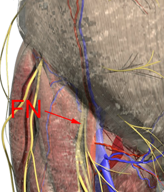
3) Nerve to pectineus (note deep to fascia iliaca) Femoral sheath (inferior projection of transversalis fascia anteriorly and iliac fascia posteriorly) 1) Lateral compartment common femoral artery and genitofemoral nerve 2) Intermediate compartment femoral vein 3) Medial compartment = femoral canal lymph node and lymphatics LandmarksAnatomy The femoral nerve is one of the major branches of the lumbar plexus The femoral nerve is consistently lateral to the femoral artery, deep to the fascia iliaca and superficial to the iliopsoas muscle The anterior approach to block the femoral nerve at the groin (inguinal region) is most commonly performed for knee surgery FA = femoral artery FN = femoral nerve FV = femoral vein


Healthy Street Anatomy Of Femoral Triangle The Femoral Triangle A Subfascial Formation Is A Triangular Landmark Useful In Dissection And In Understanding Relationships In The Groin In Living People It Appears


Femoral Triangle Sketchy Medicine


Consent Femoral Endarterectomy Teachmesurgery


S F Physical Exam Flashcards Quizlet


Dissector Answers Anterior Medial Thigh


1


Deep Artery Of The Thigh Thorax Coronal Plane Femoral Artery Lung Anatomy Png Pngegg


Femoral Artery Vein Nerve Anatomy


Close Proximity Of The Femoral Nerve Femoral Artery And Femoral Vein To The Acetabular Retractor Trialexhibits Inc


Instant Anatomy Lower Limb Vessels Arteries Femoral Artery


Femoral Artery Wikipedia
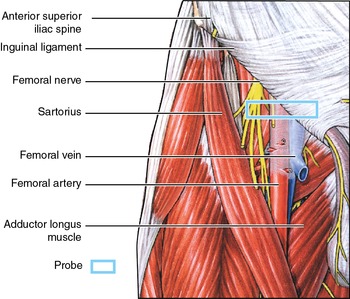


Femoral Vein Sonoanatomy For Anaesthetists


Www Vbu Ac In Resources Assets Img Dept Physio Lecture Notes On Femoral Triangle By Dr Sanjeev Kumar Pdf


Femoral Region Gastrointestinal Medbullets Step 1


Illustration Of The Case Fa Femoral Artery Fv Femoral Vein Iea Download Scientific Diagram
:background_color(FFFFFF):format(jpeg)/images/library/11105/165_blood_vessels+nerves_thigh_ventral.png)


Lower Extremities Arteries And Nerves Anatomy Branches Kenhub


Untitled Document


Thigh Knee And Popliteal Fossa Knowledge Amboss
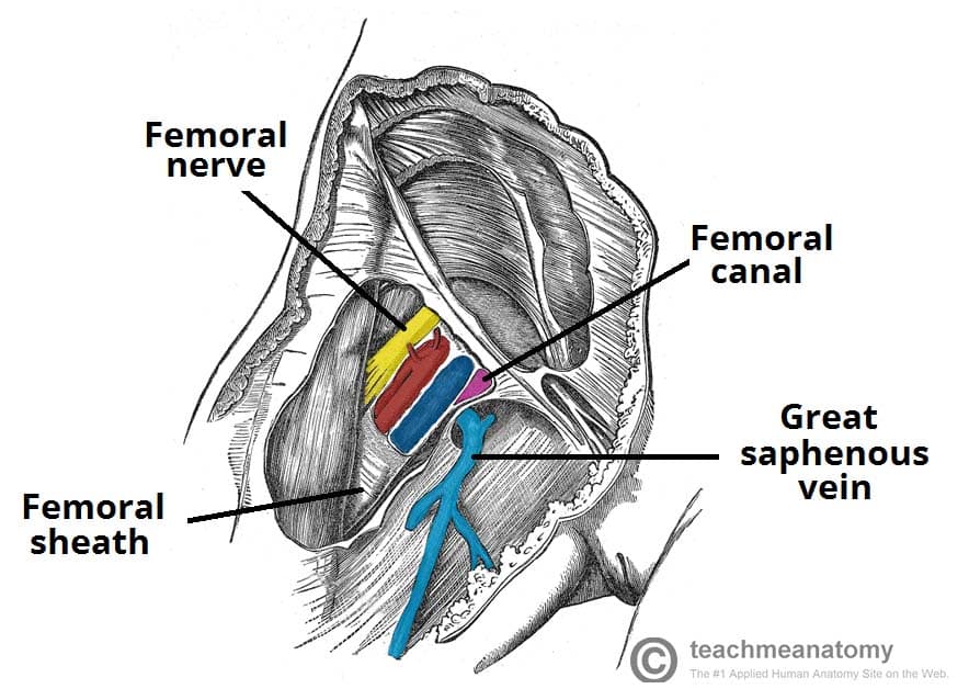


The Femoral Triangle Borders Contents Teachmeanatomy
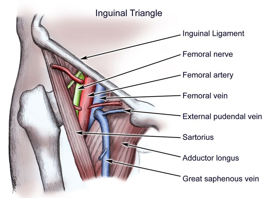


Femoral Triangle Borders Contents And Clinical Importance Medical Junction


Femoral Artery Png Images Pngwing
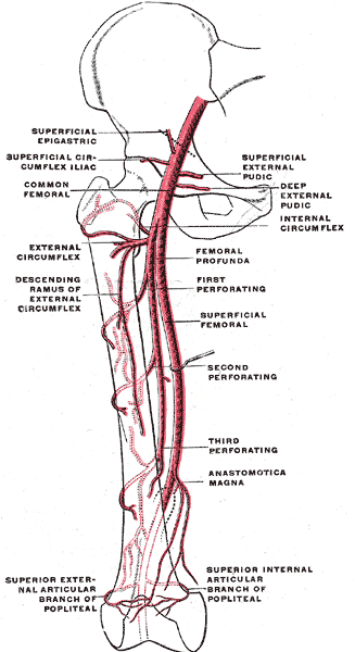


Femoral Artery Anatomy Mcq Pg Blazer


Anterior Thigh


Medical Addicts Info The Femoral Triangle Of Scarpa The Femoral Triangle Of Scarpa Is An Anatomical Region Of The Upper Inner Human Thigh Boundaries It Is Bounded By Superiorly The Inguinal


Femoral Artery Prohealthsys
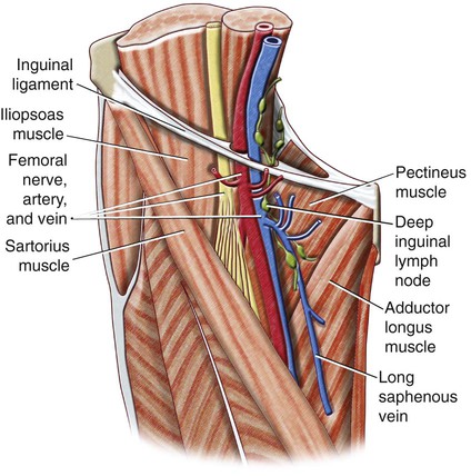


Anatomy Of The Lower Limb Radiology Key
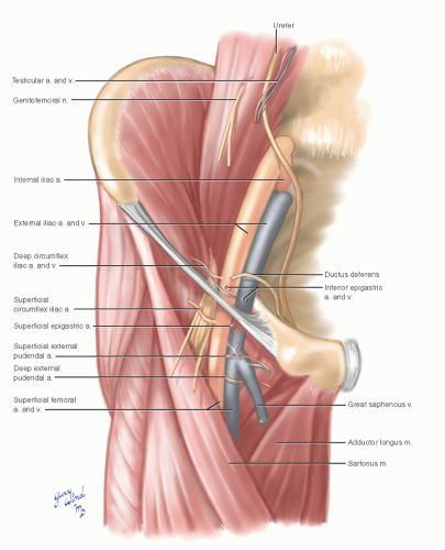


Common Femoral Artery Basicmedical Key


Femoral Vein Anatomy Aliem


Exposure Of The Common Femoral Artery And Vein Clinical Gate
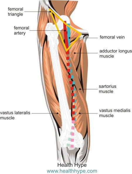


Femoral Blood Vessels Artery And Vein Anatomy Pictures Healthhype Com


Q Tbn And9gcstcjh Ewbwyp8dlquikwmsjmlypr8gcnjy4xfduhjfgvmmduxo Usqp Cau


Hip And Thigh Arteries Veins And Nerves Preview Human Anatomy Kenhub Youtube


Femoral Triangle Wikipedia


ɹǝʇlnoԁ Piʌɐᗡ 𝔹𝕖 𝕜𝕚𝕟𝕕 Anatomy Of The Femoral Triangle 1 Femoral Artery 2 Femoral Nerve 3 Femoral Vein 4 Anterior Superior Iliac Spine 5 Inguinal Ligament 6


Femoral Artery Anatomy


Jaypeedigital Ebook Reader
/GettyImages-87302280-83604c7a3ca84315a84304a002377404.jpg)


Femoral Vein Anatomy Function And Significance
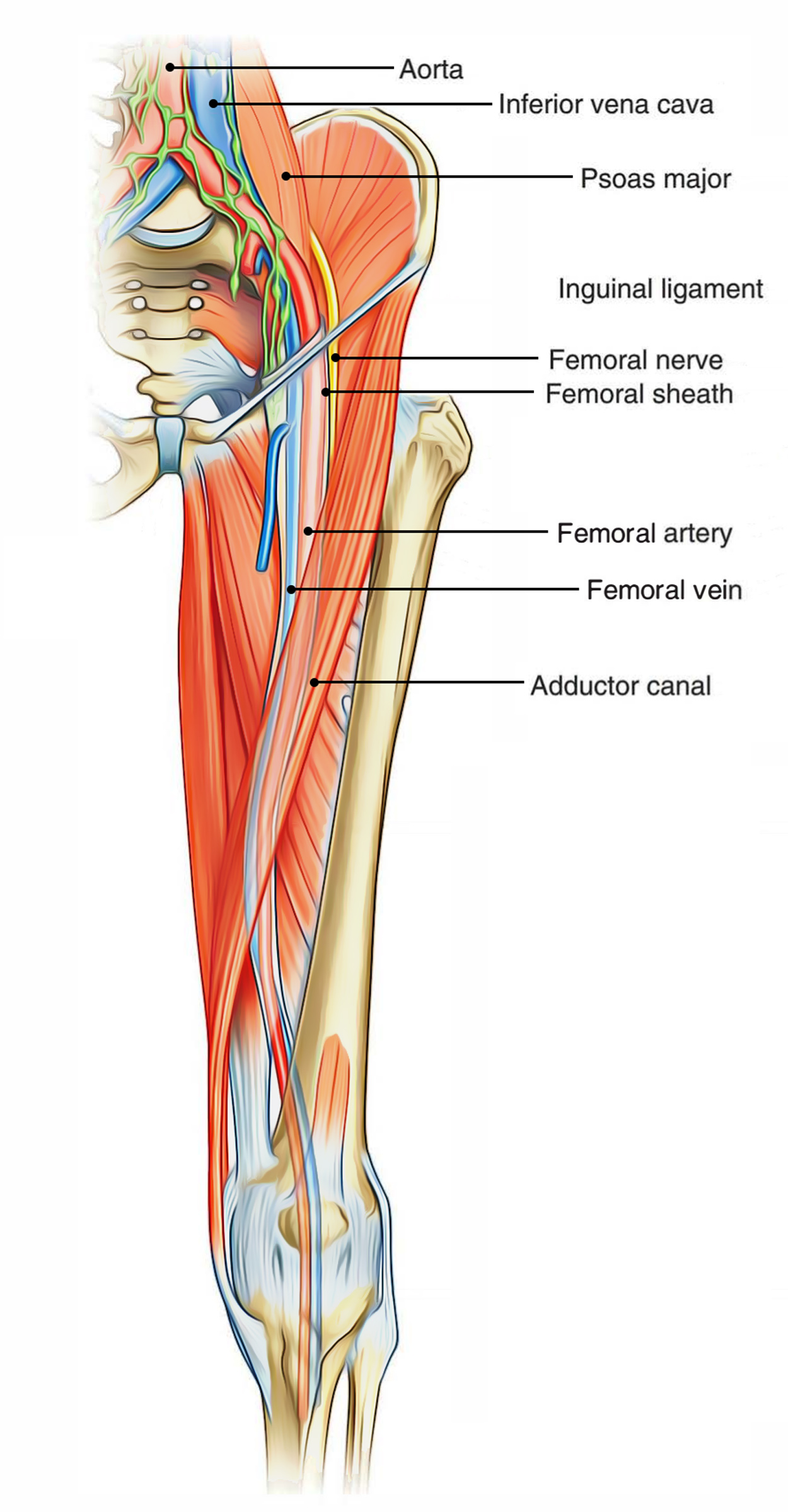


Easy Notes On Femoral Triangle Learn In Just 4 Minutes Earth S Lab
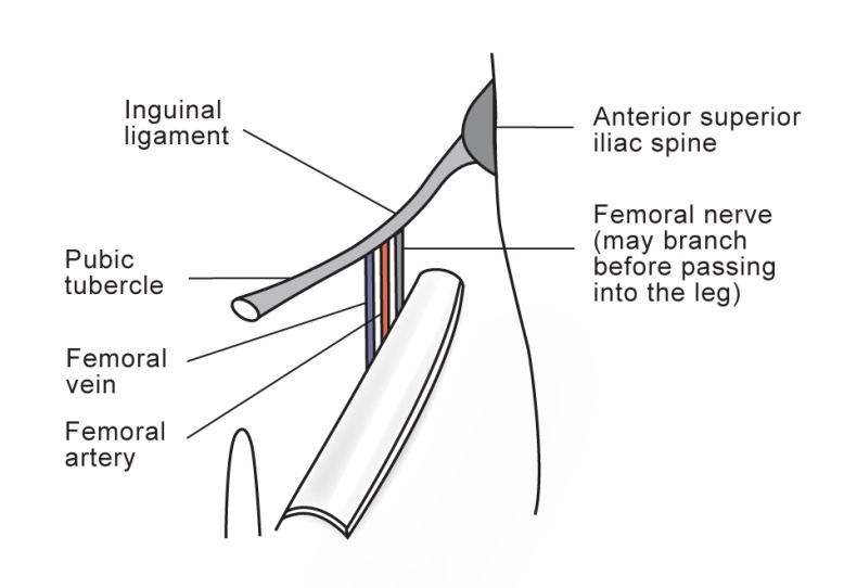


Femoral Nerve Block


Aiimsrishikesh Edu In Documents Femoral Triangle Rashmi Final 2 Pdf


Anatomy Of The Femoral Nerve Artery And Vein Medical Illustration


An Unusual Case Femoral Artery Compression


Lower Limb Anatomy The Femoral Triangle Ponder Med
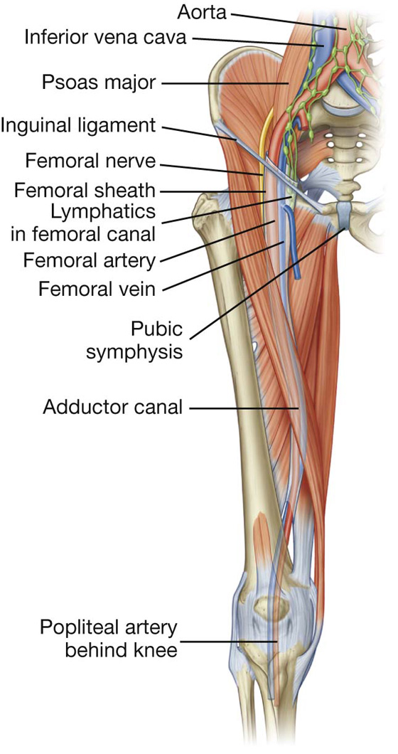


Lower Limb Basicmedical Key
/CloseupoflegwhileexercisingPeterDazeleyGettyImages-bf452734667d45ae8756ef7286e24cfd.jpg)


Femoral Artery Anatomy Function And Significance


Anatomy Of The Femoral Triangle Orthopaedicprinciples Com
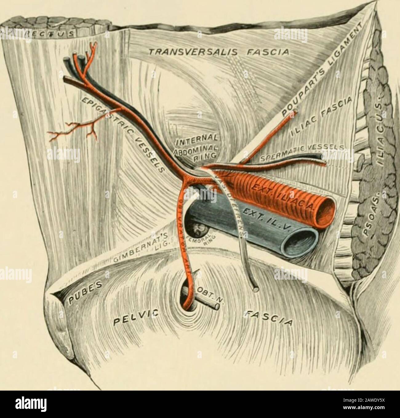


Operative Surgery Femoral Artery H Femoral Vein I Sheath Of Femoral Vessels J Saphenous Vein The Stricture Be Divided Agreeably To Directions Often Given Parallel Withthe Course Of The Epigastric Vessels Or Even


Anterior Thigh


Femoral Artery Wikiwand


Blended Simplesynapse Com Wp Content Uploads 18 08 L12 Thigh View Pdf


Sem 2 Pelvic Limb Vessels And Nerves Flashcards Quizlet


1


Http Ksumsc Com Download Center Archive 1st 436 2 musculoskeletal block Males Anatomy 17 Vasculature of lower limb Pdf


Figure 2 A Schematic Demonstrating A Coronal View Of Normal Inguinal Anatomy Schematic Showing The Relationship Of The Artery Vein And Nerve Femoral Nerve Paralysis Following Open Inguinal Hernia Repair


Special Anatomical Regions Advanced Anatomy 2nd Ed
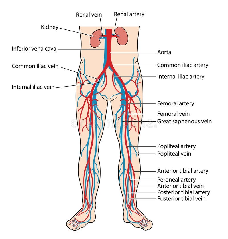


Blood Vessels Of The Lower Body Stock Vector Illustration Of Arteries Vein


Anatomy Bony Pelvis And Lower Limb Saphenous Nerve Artery And Vein Article
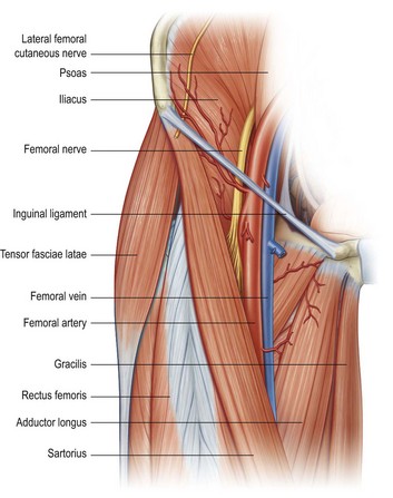


Limb Blocks Anesthesia Key


Femoral Triangle Dr Bindhu S Objectives At The


Figure Inguinal Region Inferior Epigastric Artery Statpearls Ncbi Bookshelf


Abnormalities Of The Saphenous Nerve At The Ankle Radiology Key
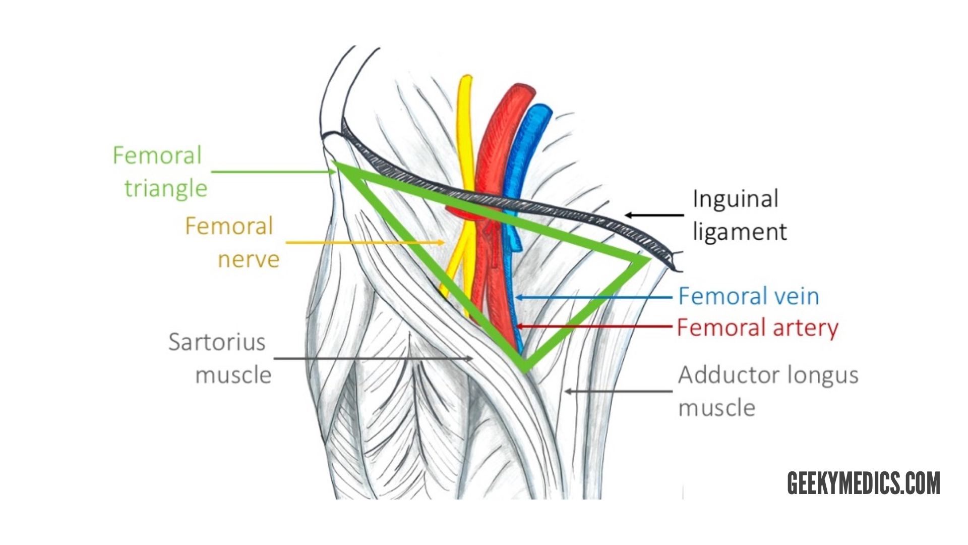


Arterial Supply Of The Thigh And Gluteal Region Geeky Medics


Focus On Venous Embryogenesis Of The Human Lower Limbs Servier Phlebolymphologyservier Phlebolymphology


The Femoral Triangle And Exposure Of The Femoral Artery Surgery Oxford International Edition


624 Femoral Artery High Res Illustrations Getty Images
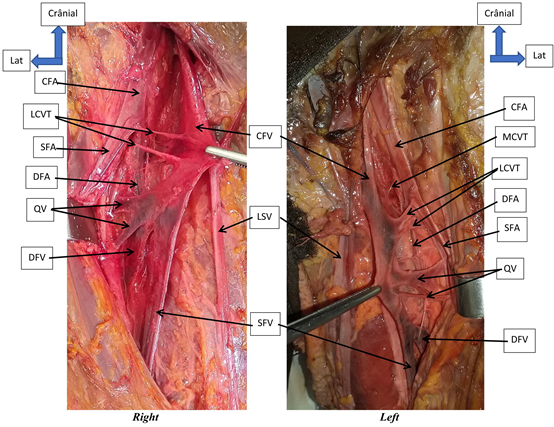


Anatomic Dissection Of The Femoral Vein At The Bamako Anatomy Laboratory
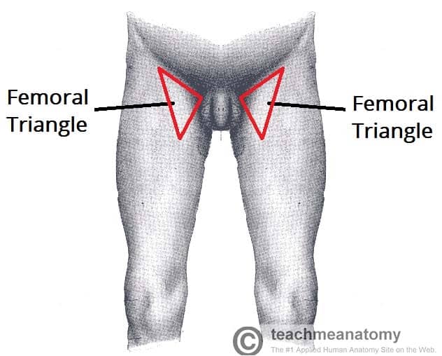


The Femoral Triangle Borders Contents Teachmeanatomy


Regional Anatomy Of A Rabbit S Thigh The Femoral Artery And Vein Download Scientific Diagram
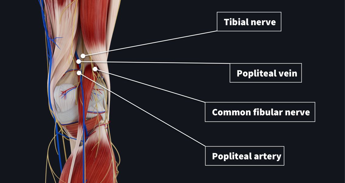


The Popliteal Fossa Complete Anatomy
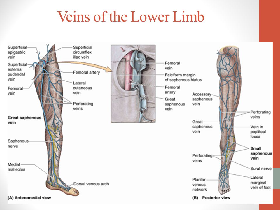


Arteries Of Leg And Foot Veins Lymphatics Of The Lower Limb Science Online


Safe And Smart Dry Needling Round Three
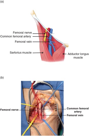


Femoral Artery Injuries Chapter 35 Atlas Of Surgical Techniques In Trauma
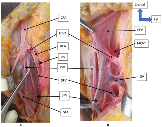


Anatomic Dissection Of The Femoral Vein At The Bamako Anatomy Laboratory


1


No comments:
Post a Comment